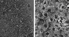
The left panel shows a raw image of Xenopus embryo tissue. This tissue is well-known to be extremely scattering, which is evident by the fact that we cannot resolve any structures in the raw image. The right panel shows the result of our computational scatter-correction method, which drastically improves imaging capability. After scatter-correction, cellular boundaries, nuclei, and yolk platelets can be clearly identified with subcellular resolution.